Please wait...
Nepřihlášený uživatel
home
iduzel: 67971
idvazba: 82273
šablona: stranka
čas: 2.5.2024 16:34:03
verze: 5351
uzivatel:
remoteAPIs:
branch: trunk
Server: 147.33.89.153
Obnovit | RAW
iduzel: 67971
idvazba: 82273
---Nová url--- (newurl_...)
domena: 'nivb.vscht.cz'
jazyk: 'en'
url: '/home/flaviviry'
iduzel: 67971
path: 8549/67607/67653/67658/67659/67971
CMS: Odkaz na newurlCMS
branch: trunk
Obnovit | RAW

Flaviviruses

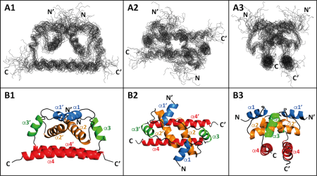
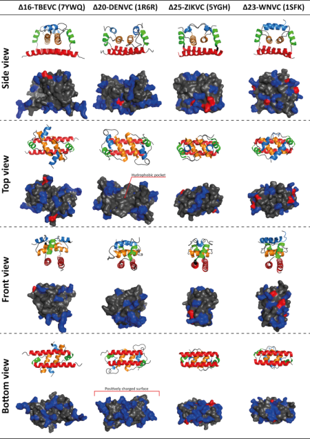
Flaviviruses such as Dengue virus (DENV), West Nile virus (WNV), Zika virus (ZIKV), Yellow fever virus (YFV), and Tick-borne encephalitis virus (TBEV) are important human pathogens belonging to the arthropod-borne viruses with a worldwide impact on human and animal health. Despite the availability of TBEV vaccine, there is still a need for tick-borne encephalitis treatment. Our research is focused on one of the promising targets for such antivirals, a flaviviral capsid (C) protein. The C protein triggers the flaviviral assembly, most probably by simultaneous binding the viral genomic RNA and ER lipid bilayer. Based on our recent publication reporting the NMR structure of TBEV C protein, we focused on the identification of the regions/residues of TBEV C involved in lipid-membrane binding and viral genomic RNA interactions. The obtained data should significantly expand our knowledge of the mechanism and specificity of C protein in the process of viral RNA packaging during TBEV infection and thus contribute to the development of targeted therapy.

A 3D structure of Δ16-TBEVC homodimer based on measured NMR spectroscopy and hydrogen-deuterium exchange mass spectrometry data. A) Superposition over the backbone heavy atoms of helices α2–α4 of the 20 dimeric Δ16-TBEVC structures represented as lines of the main chain. B) The average structure of the Δ16-TBEVC homodimer represented as a cartoon. 1—side view, 2—top view, and 3—front view. The helices are marked as α1–α4.

Comparison of homodimeric C protein structures of Tick-borne encephalitis virus (Δ16-TBEVC), Dengue virus (Δ20-DENVC), Zika virus (Δ25-ZIKVC) and West Nile virus (Δ23-WNVC). The first row of each view represents the protein structures in the cartoon representation. Blue—α1, orange—α2, green—α3, and red—α4. The second row of each view represents the protein structures with the shown surface. Positive residues are highlighted in blue, negative residues in red, and the remaining residues in gray. For Δ20-DENVC, a hydrophobic pocket (top view), which is thought to be involved in interaction with the endoplasmic reticulum membrane but is not as clearly defined in the other structures, and a positively charged surface (bottom view), which is thought to be responsible for viral genomic RNA binding and is common to all structures, are indicated.

Updated: 20.1.2023 12:44, Author: Jan Prchal

UCT Prague
Technická 5
166 28 Prague 6 – Dejvice
Identification No.: 60461373
VAT: CZ60461373

Czech Post certified digital mail code: sp4j9ch

Copyright: UCT Prague
Information provided by the Department of International Relations and the Department of Communication. Technical support by the Computing Centre.