Prosím čekejte...
Nepřihlášený uživatel
logo VŠCHT
 → Úvodní stránka → Retroviry
iduzel: 67737
idvazba: 81922
šablona: stranka
čas: 27.4.2024 05:58:39
verze: 5378
uzivatel:
remoteAPIs:
branch: trunk
Server: 147.33.89.150
Obnovit | RAW
iduzel: 67737
idvazba: 81922
---Nová url--- (newurl_...)
domena: 'nivb.vscht.cz'
jazyk: 'cs'
url: '/home/retroviry'
iduzel: 67737
path: 8549/67607/67608/67613/67614/67737
CMS: Odkaz na newurlCMS
branch: trunk
Obnovit | RAW

Retroviry

Rozpad retrovirového core po vstupu do hostitelské buňky je oblastí velkého vědeckého zájmu. Nedávné zjištění, že intaktní kapsida HIV-1 prostupuje jadernými póry do jádra infikovaných buněk, vrhlo na tento proces zcela nové světlo. Kombinací analýzy cryoEM, MS a iCLIP zkoumáme několik aspektů procesu disociace retrovirového core, včetně zapojení hostitelských kofaktorů.

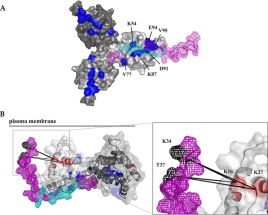
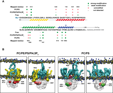
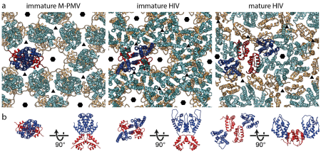
Zajímá nás také maturace, neboli štěpení retrovirového strukturního polyproteinu Gag, jejíž přesný postup je zcela kritický pro správné vytvoření zralé, plně infekční virové částice. Věnujeme se jednak poslednímu kroku maturace HIV-1, tedy odštěpení krátkého peptidu (SP1) z C-konce kapsidového proteinu (CA). Tento krok je inhibován maturačními inhibitory, jejichž nové deriváty testujeme kombinací buněčných a in vitro metod. Další oblastí našeho zájmu je také první krok samotné maturace Mason-Pfizerova opičího viru (M-PMV) a s ním spojené strukturní změny, které tento proces reorganizace retrovirové částice doprovázejí. Naše nová strukturní data ukázují, že interakce matrixové domény (MA) M-PMV s plazmatickou membránou vedou k expozici kotranslačně připojeného myristoylu, a tato změna konformace následně významně ovlivňuje počáteční krok maturace, a může tak představovat kontrolní prvek v sekvenčním štěpení polyproteinu Gag.

 Model of the CT complex with a trimeric MA. (A) MA-CT38 complex calculated in HADDOCK based on NMR results. (B) Model of the MA-CT complex created based on HADDOCK calculation (the C-terminal part of the CT38) and the results of XL-MS (the N-terminal part of the CT), where the N-terminal part of the CT sequence was twisted around the MA to meet the restrains determined by identified cross-links. Trimeric MA (30) is shown in the surface representation (A) or in the cartoon representation with a transparent surface (B), and single MA monomers are colored in different shades of gray. The MA residues identified by NMR spectroscopy as interacting with the CT38 are colored blue, and the MA residues forming cross-links with the CT38 residues are colored red. CT38 is shown in ribbon with mesh surface representation colored violet. The CT38 residues identified by NMR as interacting with the MA are colored, cyan and the CT38 residues forming cross-links with MA residues are colored black. The cross-links are displayed as dashed lines. The plasma membrane is depicted as a full line to express the orientation of the MA-CT38 complex on the membrane.Schematic model of the organization of MA and CT trimers at the membrane (a view from the inner side of the membrane). MA trimers and CT38 molecules are shown in surface representation. Single MA monomers are colored in different shades of gray, and CT38 is colored violet. Six MA molecules of individual MA trimers form a hexameric structure, with three MA trimers in complex with three individual CT38 domains of the M-PMV TM Env trimer anchored in the plasma membrane.

Surface mapping of Mouse mammary tumor virus MA  A) Interactions in the sequence of MMTV MA bound to liposomes mimicking phospholipid composition of inner leaflet of plasma membrane containing PC, PE, PS, PI4,5P2 in molar ratio of 45:45:5:5 (PC/PE/PS/PI4,5P2), and protein bound to liposomes comprised of PC and PS in molar ratio of 66:34 (PC/PS).  B) Residues of MMTV MA visualized in the snapshots obtained by GC-MD simulations. The snapshots are presented in to different side-views. Accessible residues are colored in green, inaccessible residues in red, myristoyl in orange, PS in cyan and PI4,5P2 in yellow.  Junkova, P et al., Differences and commonalities in plasma membrane recruitment of the two morphogenetically distinct retroviruses HIV-1 and MMTV. J. Biol. Chem. 2020 Jun 26;295(26):8819-8833. doi: 10.1074/jbc.RA119.011991. Epub 2020 May 8.a) Scheme of used constructs; electrophoretic verification of purity of bacterially produced proteins; TEM analysis of in vitro assembled particles; structures of used polyanions  b) Assembly efficiency of HIV-1 d16-99MACASP1NCSP2 wt protein in the presence of polyanions at indicated protein hexamer : PA ratios.  c) Fluorescence emission curves demonstrating the effect of polyanions on the kinetics of tqON degradation (the colors of the curves corresponding to the polyanions are the same as those in panel b)  Dostálková A. et al., In vitro quantification of the effects of IP6 and other small polyanions on immature HIV-1 particle assembly and core stability. J Virol. (2020) 94 (20): e00991-20, doi: 10.1128/JVI.00991-20.Colocalization of M-PMV transmembrane domain of mCherry labeled Env glycoprotein (red) with Golgi marker (green) in COS-1 cells. The samples were imaged with spinning disk confocal microscope (Andor) at magnification 600x   Grznárová Prokšová P. et al., Mason-Pfizer Monkey Virus Envelope Glycoprotein Cycling and Its Vesicular Co-Transport with Immature Particles. Viruses (2018), 10(10), 575; doi:10.3390/v10100575a) The arrangement of the CA lattices in immature M-PMV and immature and mature HIV-1 particles.  CA-NTD and CA-CTD domains - in cyan/blue and orange/red, respectively.  b) Top and orthogonal views of the structures of CA dimers from the lattices.  Schur F. et al., The structure of the immature HIV-1 capsid in intact virus particles at 8.8 Å resolution. Nature (2015) 517, 505–508, doi:10.1038/nature13838

Aktualizováno: 20.1.2023 13:05, Autor: Jan Prchal

VŠCHT Praha
Technická 5
166 28 Praha 6 – Dejvice
IČ: 60461373
DIČ: CZ60461373

Datová schránka: sp4j9ch

Copyright VŠCHT Praha
Za informace odpovídá Oddělení komunikace, technický správce Výpočetní centrum

VŠCHT Praha
na sociálních sítích
zobrazit plnou verzi